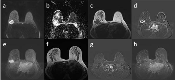
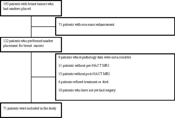
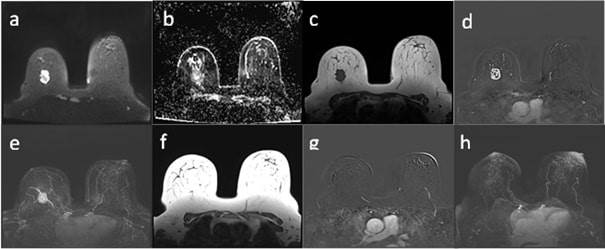
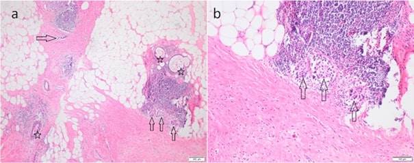
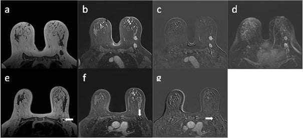
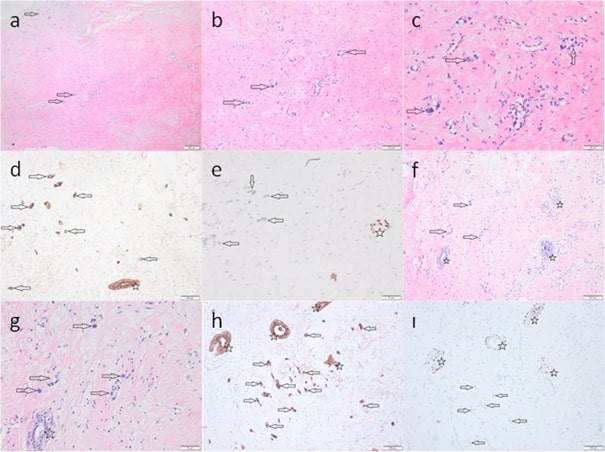
Objective: This study investigated the ability of dynamic breast magnetic resonance imaging (MRI) to predict pathological complete response (pCR) after neoadjuvant chemotherapy (NACT) in locally advanced breast cancer with marker placement and compared MRI findings with the gold standard postoperative pathology. Methods: We retrospectively evaluated Dynamic Contrast-Enhanced MRI exams of 85 breast masses in 71 NACT patients with ultrasonography-guided marker placement (2020-2023). Two radiologists, blinded to pathology, assessed the radiologic treatment response (rR) on MRI using RECIST 1.1. Pathological evaluation defined pCR as absence of invasive carcinoma. Statistical analysis provided Kappa, sensitivity, specificity, PPV, and NPV. Results: Post-NACT MRI showed 52.9% radiological complete responce (rCR), while pathology confirmed 45.2% pCR. Strong concordance between rR and pR was observed (p <0.001, ĸ = 0.740). MRI’s diagnostic performance yielded 80% sensitivity, 94.7% specificity, 95% positive predictive value (PPV) and 80% negative predictive value (NPV). False negatives related to microscopic tumors, docetaxel effects, artifacts, low histological grade, and luminal subtype. False positives linked to HER2 positivity, granulation, and fibrosis. Conclusion: MRI effectively correlates with pR post-NACT in marker-placed breast cancers, showing high specificity. For patients with MRI-evaluated complete response, pCR might be confirmed non-surgically by vacuum-assisted biopsy, though larger, long-term studies are needed for validation.
Conflict of interest : The authors declare that they have no conflict of interest.
Mevlana Tıp Bilimleri 2026, Vol6, Issue1 E-ISSN: 2757-976X
Received : , Accepted : , Published Online :
References
1. Rüland AM, Hagemann F, Reinisch M, Holtschmidt J, Kümmel A, DIttmer-Grabowski C, et al. Using a New Marker Clip System in Breast Cancer: Tumark Vision® Clip- Feasibility Testing in Everyday Clinical Practice. Breast Care. 2018;13(2):116–20.
2. Kanyılmaz G, Aktan M at al. Meme Kanserinde 5 Yıllık Tedavi Sonuçlarımız ve Prognostik Faktörler: Tek Merkez Deneyimi. Selçuk tıp Dergisi. 2017;33(1):5–9.
3. Whitman GJ, Iyer RB, Reeve CJ, Patel PR, Phelps MJ, Pusztai L. Assessment of response to neoadjuvant chemotherapy in breast cancer: Imaging considerations. Semin Breast Dis. 2004;7(2):61–74.
4. Soliman AH, Osman AM. Cost-effectiveness of ultrasound-guided surgical clips placement for breast cancer localization prior to neoadjuvant chemotherapy. Egyptian Journal of Radiology and Nuclear Medicine [Internet]. 2018;49(4):1163–8. Available from: https://doi.org/10.1016/j.ejrnm.2018.06.010
5. Youn I, Choi SH, Kook SH, Choi YJ, Park CH, Park YL, et al. Ultrasonography-guided surgical clip placement for tumor localization in patients undergoing neoadjuvant chemotherapy for breast cancer. J Breast Cancer. 2015;18(1):44–9.
6. Hossam A, El-Badrawy A, Khater A, Setit A, Roshdy S, Abdelwahab K, et al. The Evaluation of a Cost-Effective Method for Tumour Marking Prior to Neo-Adjuvant Chemotherapy Using Silver Rods. Eur J Breast Health. 2023;19(1):99–105.
7. Zhang X, Wang D, Liu Z, Wang Z, Li Q, Xu H, et al. The diagnostic accuracy of magnetic resonance imaging in predicting pathologic complete response after neoadjuvant chemotherapy in patients with different molecular subtypes of breast cancer. Quant Imaging Med Surg. 2020;10(1):197–210.
8. Romeo V, Accardo G, Perillo T, Basso L, Garbino N, Nicolai E, et al. Assessment and prediction of response to neoadjuvant chemotherapy in breast cancer: A comparison of imaging modalities and future perspectives. Cancers (Basel). 2021;13(14):1–18.
9. Amin MB, Edge SB, Greene FL, Byrd DR, Brookland RK, Washington MK, et al. AJCC cancer staging manual. Vol. 1024. Springer; 2017.
10. Kataoka A, Sawaki M, Horisawa N, Kotani H, Yoshimura A, Hattori M, et al. The Absence of Cancer in the Location of a Breast Tissue Marker After Neoadjuvant Chemotherapy may Predict Pathological Complete Response with High Accuracy: Results from a Phase II Trial. Ann Surg Oncol [Internet]. 2023;30(6):3224–32. Available from: https://doi.org/10.1245/s10434-023-13199-8
11. Aebi S, Karlsson P, Wapnir IL. Locally advanced breast cancer. Breast [Internet]. 2022;62: S58–62. Available from: https://doi.org/10.1016/j.breast.2021.12.011
12. Parra RFD Van, Kuerer HM. Selective elimination of breast cancer surgery in exceptional responders: historical perspective and current trials. Breast Cancer Research [Internet]. 2016;1–8. Available from: http://dx.doi.org/10.1186/s13058-016-0684-6
13. Shalaby LASED, Khallaf ES el din, Moussa MM. Clip and wire localization of locally advanced malignant breast masses in patients undergoing neoadjuvant chemotherapy and breast conservation therapy. Egyptian Journal of Radiology and Nuclear Medicine. 2019;50(1).
14. Mitzi IIM, Freire G, Carrara A, Scapulatempo-neto IC, Lucas II, Abraha F, et al. Breast-conserving surgery in locally advanced breast cancer submitted to neoadjuvant chemotherapy. Safety and effectiveness based on ipsilateral breast tumor recurrence and long-term follow-up. 2017;134–42.
15. Clouth B, Chandrasekharan S, Inwang R, Smith S, Davidson N, Sauven P. The surgical management of patients who achieve a complete pathological response after primary chemotherapy for locally advanced breast cancer. 2007; 33:961–6.
16. Sanli AN, Turan B, Tekcan Sanli DE, Karaca I, Altundag MK, Aydogan F. Neoadjuvant response stratification based on complete, partial, and no response in HR-positive/HER2-positive breast cancer. Breast Cancer Res Treat [Internet]. 2025;(August). Available from: https://doi.org/10.1007/s10549-025-07812-5
17. Popa E, Croitoru A, Cristian D, Jitea N, Scaunasu R, Aldea C, et al. Surgical features after neoadjuvant treatment for breast cancer. Chirurgia (Romania). 2021;116(2):193–200.
18. Wolmark N, Wang J, Mamounas E, Bryant J, Fisher B. Preoperative Chemotherapy in Patients With Operable Breast Cancer: Nine-Year Results From National Surgical Adjuvant Breast and Bowel Project B-18 AND. 2001;15212(30).
19. Wong SM, Santos JDL, Basik M. Eliminating Surgery in Early-Stage Breast Cancer: Pipe-Dream or Worthy Consideration in Selected Patients? 2017;148–55.
20. Heil J, Ku S, Schaefgen B, Paepke S, Thomssen C, Rauch G, et al. Diagnosis of pathological complete response to neoadjuvant chemotherapy in breast cancer by minimal invasive biopsy techniques. 2015;(October):1565–70.
21. Noordaa MEM Van Der, Duijnhoven FH Van, Loo CE, Werkhoven E Van, Vijver KK Van De, Wiersma T, et al. Identifying pathologic complete response of the breast after neoadjuvant systemic therapy with ultrasound guided biopsy to eventually omit surgery: study design and feasibility of the MICRA trial (Minimally Invasive Complete Re. The Breast. 2018;
22. Mauriac L, Macgrogan G, Avril A, Durand M, Floquet A, Debled M, et al. Original article Neoadjuvant chemotherapy for operable breast carcinoma larger than 3 cm: A unicentre randomized trial with a 124-month median follow-up. 1999;47–52.
23. Ring BA, Webb A, Ashley S, Allum WH, Ebbs S, Gui G, et al. Following Neoadjuvant Chemotherapy for Early Breast Cancer? 2015;21(24):4540–5.
24. Aveau CAD, Avignoni ALS, Oumya S, Nane AB, Endale MID, Ranc F, et al. IS RADIOTHERAPY AN OPTION FOR EARLY BREAST CANCERS WITH COMPLETE CLINICAL RESPONSE AFTER NEOADJUVANT CHEMOTHERAPY? ¸ OIS C AMPANA, M. D., * Patients characteristics. 2011;79(5):1452–9.
25. Matthias H, Clémence B, Electra S, Thanh L, Poletti Pierre-Alexandre P, Alexandre B, et al. Diagnostic precision of breast MRI in prediction of pathological complete response: Is it influenced by the presence of metallic markers? Eur J Radiol. 2022;154(July).
26. Talec H, Aubé C, Guerin-Charbonnel C, Berge P. The use of a clip prior to neoadjuvant chemotherapy for breast cancer with microcalcifications may not always be required. Breast Cancer Res Treat [Internet]. 2025;209(3):585–93. Available from: https://doi.org/10.1007/s10549-024-07517-1
27. Kwon M ri, Chu J, Kook SH, Kim EY. Factors associated with radiologic-pathologic discordance in magnetic resonance imaging after neoadjuvant chemotherapy for breast cancer. Clin Imaging [Internet]. 2022;89(April):1–9. Available from: https://doi.org/10.1016/j.clinimag.2022.05.002
28. Moon H, Han W, Lee JW, Ko E, Kim E, Yu J, et al. Age and HER2 expression status affect MRI accuracy in predicting residual tumor extent after neo-adjuvant systemic treatment. 2009;(January):636–41.
29. Kim J, Han BK, Ko EY, Ko ES, Choi JS, Park KW. Prediction of pathologic complete response on MRI in patients with breast cancer receiving neoadjuvant chemotherapy according to molecular subtypes. Eur Radiol [Internet]. 2022;32(6):4056–66. Available from: https://doi.org/10.1007/s00330-021-08461-0
30. Turnbull LW. Dynamic contrast-enhanced MRI in the diagnosis and management of breast cancer. 2009;(March 2008):28–39.
31. Morrow M, Waters J, Morris E. Breast Cancer 1 MRI for breast cancer screening, diagnosis, and treatment. The Lancet [Internet]. 2011;378(9805):1804–11. Available from: http://dx.doi.org/10.1016/S0140-6736(11)61350-0
32. Richter H, Hennigs A, Schaefgen B, Hahn M, Blohmer JU, Kümmel S, et al. Is Breast Surgery Necessary for Breast Carcinoma in Complete Remission Following Neoadjuvant Chemotherapy? Ist eine Operation der Brust bei Komplettremission nach neo- adjuvanter Chemotherapie des Mammakarzinoms notwendig? Authors Historical Studies on. 2018;48–53.
33. Choi WJ, Kim HH, Cha JH, Shin HJ, Chae EY, Yoon GY. Complete response on MR imaging after neoadjuvant chemotherapy in breast cancer patients: Factors of radiologic-pathologic discordance. Eur J Radiol [Internet]. 2019;118(March):114–21. Available from: https://doi.org/10.1016/j.ejrad.2019.06.017
34. Ko ES, Han B kyung, Kim RB, Ko EY. Analysis of Factors that Influence the Accuracy of Magnetic Resonance Imaging for Predicting Response after Neoadjuvant Chemotherapy in Locally Advanced Breast Cancer. 2013;2562–8.
35. Negrão EMS, Souza JA, Marques EF, Bitencourt AGV. Breast cancer phenotype influences MRI response evaluation after neoadjuvant chemotherapy. Eur J Radiol [Internet]. 2019;120(September):108701. Available from: https://doi.org/10.1016/j.ejrad.2019.108701



 Keywords